By Dr. John R. Mishock, PT, DPT, DC
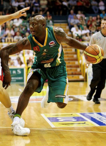
An acute ankle sprain is the most common lower limb injury accounting for up to 40% of all sports-related injuries. The highest incidence of injury occurs in basketball (41.1%), American football (9.3%), and soccer (7.9%). (Vuurberg et al. Br j Sports Med, 2018, (McKay et al Br J Sports Med, 2001). Ankle sprains not only limit playing time during the acute phase but they also can become recurrent, chronic and potentially debilitating with 70% of individuals never fully recovering. (Braun et al. Arch Fam Med, 1999; Hubbard Open Access J, 2010)
What is injured during an ankle sprain?
Almost 85% of ankle sprains involve the outside ankle ligaments (3 lateral ligaments: anterior talofibular, calcaneofibular, or posterior talofibular). The mechanism of injury is an inversion movement in which the foot is forcefully turned inward, often during a change of direction movement or stepping on another athlete’s foot. In most cases, (65%), isolated injury occurs in the anterior talofibular ligament (front outside ligament). In 20%, injury of both anterior talofibular and the calcaneofibular ligaments (front and side ligaments) occurs. (Doherty et al. Sports Med, 2014)
Less than 15% of injured ankles involve the inside (medial ankle ligaments) ligaments or a “high ankle sprain”. The “high ankle sprain” is an injury to the ligamentous complex that holds the tibia and fibula together (syndismosis) located in the front lower leg (distal tibiofibular junction). (Asian et al. OA Orthop 2014)
Do I need x-rays following an acute ankle sprain?
In most cases, x-rays are not needed following an ankle sprain. The Ottawa Ankle and Foot Rules were developed to guide health care providers on the need for x-rays. (Bachmann et al. BMJ, 2003) Multiple studies have validated the Ottawa ankle and foot rules with high sensitivity of 94.6% to 100% in detecting fractures in adults and children with acute ankle injury. (Lau et al. JBJS Rec 2018; Ivins et al Am Fam Phys, 2006; Dowling et al. Acad Emerg med, 2009; Stiell et al. JAMA, 1994) Positive findings do not reveal the existing fracture but do show the need for radiography to rule out a fracture.
According to these rules, plain ankle x-rays are needed, if pain or tenderness exists to palpation of the medial or lateral malleolus (boney bumps on the inside or outside of the ankle) or an inability to walk for 4 steps immediately after injury.
If there is pain in the foot (midfoot) and an inability to walk for 4 steps just after the injury or tenderness over the foot bones (navicular bone and base of the fifth metatarsal bone) x-rays are indicated. (Bachmann et al BMJ, 2003)
MRI (magnetic resonance imaging) is not indicated in the routine investigation of acute ankle injuries due to high incidence, limited accessibility, high cost, and false-positive findings. (Gribble et al. Br J Sports Med, 2016)
How are acute ankle sprains graded?
Lateral ankle sprains are classified as grade I, II, or III, based on the severity of injury to the lateral ligaments. The clinical grading is based on the amount of swelling, pain, ecchymosis (bruising), range of motion, and stability testing.
Grade I: Mild swelling (less 0.5 cm fig 8 measurement), minimal or no point tenderness to palpation, normal weight-bearing and function, minimal range of motion loss (< 5 degrees), negative orthopedic testing (anterior drawer, talar tilt)
Grade II: Moderate swelling (greater 0.5-2 cm, fig 8 measurement), point tenderness to palpation, some loss of function with normal weight-bearing, range of motion loss (5-10 degrees), positive orthopedic testing (positive anterior drawer, negative talar tilt)
Grade III: Moderate-severe swelling (>2cm, fig 8 measurements), severe point tenderness to palpation, severe loss of function and difficulty with normal weight-bearing, range of motion loss (>10 degrees), positive orthopedic testing (positive anterior drawer and talar tilt).
Asian et al. OA Orthop, 2014)
Grade III ankle injuries are rare but have a 96% likely hood of ligament rupture. (Ivins et al. Am Fm Phys, 2006)
Do I need to immobilize the ankle in a boot following an acute ankle injury?
Immobilization of the ankle with a walking boot should be judiciously used due to the deleterious effects of immobilization. If there is a fracture of the ankle or foot, immobilization is needed to allow healing (2-4 weeks). For severe grade III ankle injuries with potential ligament tears and instability, some evidence showed that a short episode of immobilization (less than 10 days) decreases pain and swelling and improves functional outcome (Uslu et al. J Am Podiatr Med Assoc, 2015; Lamb et al. Lancet, 2009; Prado et al, Foot Ankle Int, 2014). However, immobilization should be coupled with early physical therapy treatments to promote healing and expedite functional recovery.
For the grade, I and II ankle injuries there is NO evidence for immobilization with a walking boot. In most cases the immobilization is counterproductive for healing and return to function and sport. The greater the time immobilized in walking boot the greater the potential for altered healing such as; poor ligamentous scar tissue healing strength, hastened range of motion gains, slowed swelling reduction, progressive weakness of the lower limb, and delayed functional return to sports. There is also a chance for chronic symptoms at 3 to 6 months after injury along with increased ligamentous laxity on radiographs (Ardevol et al. Surg Sports Traumatol Arthrosc, 2002)
The walking boot alters normal gait mechanics and can lead to problems in the knee, hip, or low back. If normal ambulation is an issue due to pain the short-term use of crutches could be utilized. The crutches should be used to de-load the joint while maintaining normal gait mechanics.
Should I wear an ankle brace immediately following an ankle injury?
A figure 8 lace-up ankle brace is ideal to wear following an ankle injury. This type of brace allows mobility and movement but prevents end-range motion that can re-injury the ankle ligaments. The ankle could also be taped if bracing is not available. (Kerkhoffs et al. Cochrane Database Syst Rev 2013)
Does R.I.C.E work?
Similar to many acute musculoskeletal injuries, the principles of rest, ice, compression, and elevation (RICE) are historically applied for patients with an ankle sprain. However, there is limited evidence supporting the efficacy of this approach in reducing associated symptoms following injury. (Chen et al. Curr Sports Med Rep, 2019)
Although limited evidence exists RICE makes physiological sense as it most likely would cause blood vessels in the area to vasoconstrict reducing the negative effects of sustained swelling. (Hubbard et al J Athl Train, 2004). Ice should be used for 20 minutes every two hours. An ACE bandage can be used for compression. RICE should be complemented with early physical therapy (pain free ROM and mobility exercises) along with controlled weight-bearing activities.
When should I start to walk on my injured ankle?
The sooner the athlete can begin to walk the better. Numerous studies have demonstrated that integrating early weight-bearing, walking, and movement has a positive influence on the reduction of swelling, restoration of normal range of motion, and improved function. (Kerkhoffs et al. Arch Orthop Trauma Surg, 2001; Eiff et al. Am J Sports Med, 19194; Dettori et al. Mil Med, 1994) This further refutes immobilization in a walking boot.
If needed a walking-aid device such as a crutch can be used to begin weight-bearing at the individual’s tolerance to loading of the ankle. It is important to focus on a normal gait pattern as soon as possible (heel-strike, flat-foot, toe-off).
Why is early physical therapy important in recovery?
Physical therapy can make a significant impact on recovery and should begin with in the first few days to a week following injury. Countless studies have demonstrated that early physical therapy reduces pain, increases range of motion, reduces swelling, increases strength of lower extremity, enhances motor control/balance, leads to early return of function, promotes return to sport, and prevents or minimizes re-injury. (Vuurberg et al. Br J SPorts Med; 2018; Naeem et al. Orthop Traumatol, 2015, Uslu et al. J Am Podiatr Med Assoc, 2015; Lamb et al. Lancet, 2009; Prado et al, Foot Ankle Int, 2014).
As the physical therapy progresses the individual will be functionally tested and progressed based on their individual ability to heal from injury. Over time they will be trained to meet the demands of the sport such as; sprinting ability, change-of-direction movements in all direction, first step quickness, jumping (horizontal, lateral, and vertical), and read-react sports specific skills.
How does manual THERAPY help?
Manual therapy techniques (hands on soft tissue techniques, mobilization/manipulation) have been shown to critical in the early phase of healing in reduce swelling, increase range of motion, improved weight-bearing, and increase functional return to sports and work. (Vicenzion et al. J Ortho Sports Phys Ther, 2006; Denegar et al. J Orthop Sports Phys Ther, 2002; Loudon et al. Br j Sports Med, 2014) Manual therapy enhances and strengthens scar formation to optimize ligament tensile strength. (Weerasekara et al. Arch Phys Med Rehabil, 2018)
How does exercise help?
There are many research articles supporting the early use of physical therapy and rehabilitation exercise to reduce pain and swelling, increase strength, and return the athlete to early function and sport. (Docherty et al. J Athl Train, 1998; McKeon et al. J Athl Train, 2008; Wester et al. J Orthop Sports Phys Ther, 1996; Bellows et al. J Sports Phys Ther, 2018)
Exercise therapy should be comprehensive and progressive and begin as soon as possible in pain-free functional activities. Exercises should include; gait training, range of motion activity, flexibility (stretching), resistance (strengthening), neuromuscular and proprioceptive (balance and coordination), and finally sport-specific functional exercises for return to play.
How soon can I return back to my sport?
Returning an athlete back to their sport is individualized based on the steady progression of their rehab via consistent test and re-test of their functional abilities. Each visit the athlete is assessed and progressed based on functional testing relative to their ability to perform pain free movements without risk of re-injury.
Prior to the athlete returning to their sport they will be functionally tested on their ability to reproduce the demands of the sport such as; sprinting ability, change-of-direction movements in all direction, first step quickness, jumping (horizontal, lateral, and vertical) and read-react sports specific skills.
What is the risk of re-spraining my ankle?
Unfortunately, the risk of re-sprain in the first year is as high as 70%. Physical therapy exercises that focus on balance, coordination, motor control, and functional restoration can significantly reduce the chances of re-injury. (Braun et al. Arch Fam Med, 1999; Hubbard Open Access J, 2010) Beyond this, wearing a figure 8 lace up brace will significantly reduce or eliminate the chance of re-injury.
Can ankle braces reduce ankle sprains in healthy athletes who have never had an ankle injury?
It is often thought that wearing ankle braces prophylactically (preventively in healthy individuals) will cause the ankles to become weak and stiff thus increasing the chance of injury when braces are not worn. However, scientific evidence refutes this common belief. In healthy athletes without a previous ankle injury it has been shown that ankle braces can significantly improve dynamic postural control, single-limb stability and reduced the incidence of ankle sprains. (J Sport Rehabil. 2015, AM J Sports Med 2011) In multiple studies, the prophylactic use of ankle braces in healthy athletes can reduce the incidence of ankle injury by 70% without causing weakness or limited range of motion. (Dison et al. J Sci Med Sport, 2010; Richie et al. Clin Podiatr med Surg, 2015)
Keep in mind, un-like the walking boot that eliminates ankle movement and function, the figure 8 lace-up brace allows full range of motion but prevents extremes of end range motion that damage the ankle ligaments during inversion.
Dr. Mishock is one of only a few clinicians with doctorate level degrees in both physical therapy and chiropractic in the state of Pennsylvania.
He has also authored two books; “Fundamental Training Principles: Essential Knowledge for Building the Elite Athlete“, “The Rubber Arm; Using Science to Increase Pitch Control, Improve Velocity, and Prevent Elbow and Shoulder Injury” both can be bought on Amazon.
If pain or limited function is keeping you from doing the activities you enjoy,
We can help!
Reduce pain and increase function
Call for a FREE Phone Consultation or to schedule your visit (610)327-2600.
Locations: Gilbertsville, Skippack, Phoenixville, Steiner Medical, Boyertown, Pottstown, and Limerick (inside the Spring Valley YMCA).
Appointments available 7:00am to 8:00pm, ALL locations, most days!
Saturday Appointments available
Visit our website to REQUEST AN APPOINTMENT, read informative articles, meet our physical therapy staff, and learn about our treatment philosophy.